November 2022
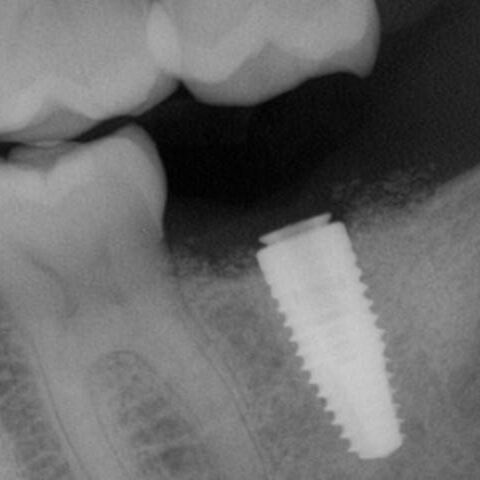
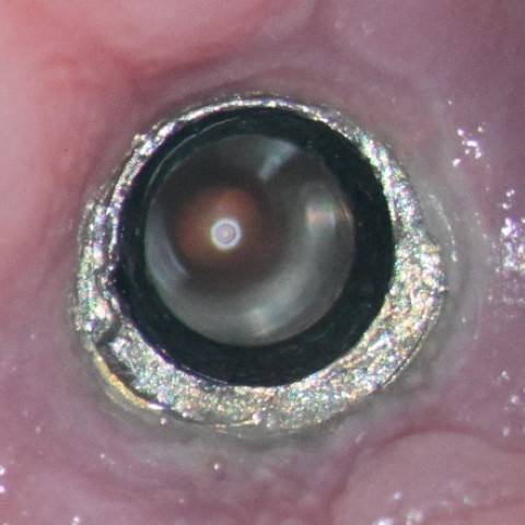
The patient presented on referral for retrieval of a fractured abutment from a 4.8 Straumann BL RC implant in the #18 site. This restoration was placed approximately 10 months ago and although the restoration was not available to examine it appeared, from the radiograph and the clinical presentation, this was a fractured TiBase abutment. There were at least two attempts to recover the abutment fragment, including needle nose pliers, Cavitron ultrasonics and endo ice. In a conversation with the Straumann rep., it was suggested this fragment might be “cold welded”. Generally, recovering RC abutment fragments is not challenging as the abutment connection is basically a slip fit joint (Straumann calls it the cross-fit connection) and above the cross-fit connection there is a top lead in bevel which is .7mm in vertical height, with a 15-degree taper. Recovering this case was no exception, and the fragment was recovered with a precision, custom through bore tool using an aggressive “wobbling” technique. Following retrieval, the implant was internally cleaned and found to be free of defects when inspected at 25x with the clinical microscope. The supplied healing abutment was placed finger tight, and the patient was referred back to the referring Dr. for restoration.
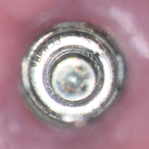
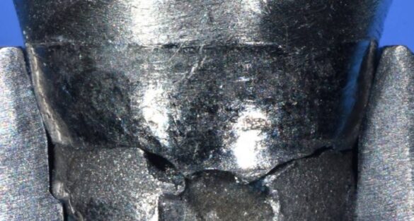
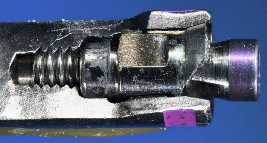
The following photographs are included to point out the mechanical “weak link” in the TiBase concept. The vertical cylinder which fractured at the base of the shoulder has a very narrow wall thickness. I have the habit of calculating the surface area of most abutment fractures I recover and the list keeps growing. Therefore, this recovered fractured abutment fragment was measured on my 14” optical comparitor in my machine shop and the area of the fracture was found to be 3.217 sq. mm. (The wall thickness measured .4mm, the outside diameter was 2.96mm, and the inside diameter of the counter bore was 2.16mm) The counter bore can be visualized in both of the photographs below and extends below the implant top. This position does decrease the cross sectional area of the cylinder by about 1.65 sq.mm. (The through bore diameter measured 1.63mm vs the 2.16mm diameter when the fracture is through the counter bore at the implant top).
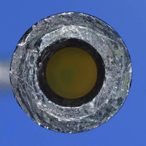
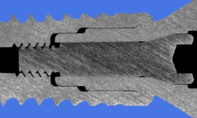
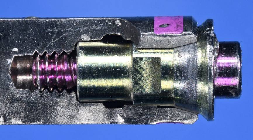
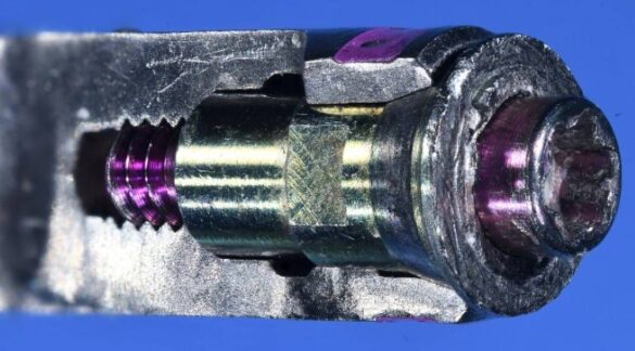
Actual fractured abutment fragment on an open analog horizontal and oblique views.
Prior to leaving the appointment the patient asked a great question as to what might be changed to avoid such a fast failure. This led into a discussion of abutment options. First, I’d like to say there is no indication this was an actual Straumann abutment, as there was no identification laser engraving on the part. I would speculate it was non-OEM. However, to illustrate the point, in the following prior photographs, another Straumann case with the abutment was laser engraved, had also failed. Arguably, a better design, but still a narrow cylinder. This case was restored in 01.20.2020 and was rescued on 06.08.21.
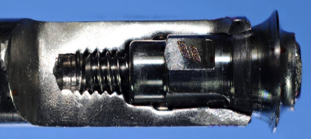
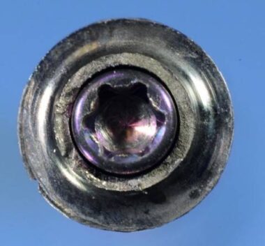
Straumann TiBase abutment with laser engraving, cylinder fracture just above the shoulder junction which was rounded to avoid creating a stress riser at the shoulder and cylinder junction, which also added material and strength. Clearly a better design but failed also in the cylinder cross section.
Therefore, I believe the choice of a custom titanium abutment would be far superior to a TiBase design because it offers a thicker wall and therefore additional strength in the vulnerable area at or just above the implant top. While it is true, I have retrieved many abutment fractures in RC implant abutments at both the implant top and in the area just below the .7mm lead in bevel in custom abutments other than TiBase designs, the failures tend to occur in years of function rather than in just months, as in this case. The two case examples below represent abutment fractures at various levels through the conical interface and represent a limitation based on interface design and size.
– Charles A. Mastrovich, DDS
