February 2021
The patient presented on referral for diagnosis and possible “retapping” of a 4.2 Implant Direct Legacy III implant in the #19 site. This implant was placed on 10.28.14 and restored on 02.16.15. According to the patient, after approximately six years in function, the abutment screw fractured. The patient was referred to an endodontist, who recovered the screw fragment over a 2+ hour appointment using ultrasonic instrumentation. Evidently, they realized there was implant thread damage, as a tap was procured from Implant Direct. It is unclear as to what the overall strategy was to correct this problem, but the end result was not successful, in that there were not enough threads to properly secure a new abutment screw. When she presented to my office the crown was loose on the implant, which had been the case for some time. The scope of this appointment was to evaluate the internal condition of the implant and assess what the best course of action should be. Therefore, the abutment screw and crown were retrieved, and the internal aspects of the implant were microscopically examined and photographed at 25x. For additional mechanical evaluation, and with the implant clean and dry, a polyvinyl impression was taken of the implant interior. Additional photographs were processed post appointment.


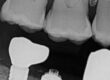
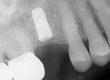
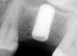
Initial preop radiographic images. Note the severe distal implant position and mesial cantilevered restoration.


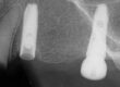
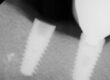
From the above images, it is clear that the thread damage is extensive, secondary to loss of the implant female threads. This was due to iatrogenic damage incured in the process of attempting to retrieve the abutment screw with ultrasonic instrumentation. The flowing damage, without sharp bur marks, is characteristic of ultrasonic use. Failure to keep the ultrasonic instrument concentric on the screw fragment can produce implant damage quite quickly. This issue can better be understood by examining a short study I have posted on our website: ULTRASONICS: Complications of Abutment Screw Retrieval Secondary to Prior Ultrasonic Attempts. This simple study illustrates the extensive damage an ultrasonic instrument can do to the internal aspects of an implant in a brief time period.
Ultrasonic instrument use is difficult, if not impossible, to control within the implant. Any recovery process should be continually verified with microscopic visualization for early correction of any eccentric diviation into the implant. Passing the original 1-72 tap through these threads did not, and will not, improve the situation. However, this case has two other glaring problems, in addition to the mechanical one described above: a biologic issue with crestal bone loss and an implant positional problem. This implant was placed to the distal lingual, for some unknown reason, which creates additional torsional loading when there is force placed on the mesial and facial of the crown. This can be easily appreciated in the preop radiographic images above and the photographs of the crown on an analog below. Torsional loading is applied force x leverage arm, and the force magnifacation is amazing as the leverage arm increases (“Physics 101”). This 3.5 TSV interface is small as compared to the size of the molar and does not protect the abutment screw well from this torsional force, even if all of the above factors were favorable. The bottom line is the implant is not a good candidate for a heroic rescue and should be explanted instead. These issues were discussed with the patient and she requested a referral to a qualified surgeon who has experence in explantation and grafting of the site.


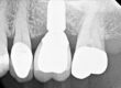
Facial view (L) and distal view (R) of the crown on the analog. The torsional loading on this implant-abutment interface is appearent.
For additional information regarding these procedures, there are additional case studies posted on our website.
Charlie Mastrovich, DDS