This redacted case study report was written as the recovery progressed outlining the thought process, clinical decisions made, unique precision tooling used and the microscope visualization necessary to successfully complete the case.
09.14.22 The patient was referred by Dr. S. for retrieval of a fractured Locator abutment screw from a Straumann 4.1 BL implant in the #3 site. This implant was placed in January of 2014 and the Locator placed in July of 2014. According to the patient, her new removable partial denture made for this attachment had excessive retention, so it was not used, and she reverted back to her older prosthesis. Upon questioning, she was unsure when this abutment fracture had occurred. This is an interesting question because if it had been several years in function, I would suspect the abutment had loosened and the fracture was secondary to lack of contact in the minimal conical connection of the RC implant. However, if the fracture had occurred very quickly there is the distinct possibility the fracture was secondary to a mismatched abutment (NC abutment in an RC implant). If an NC Locator abutment is mistakenly installed into an RC implant, it will thread in and from a clinicians viewpoint all has gone well. Unfortunately, the screw will bottom out past the implant threading before the conical interface engages so all of the installation torque becomes 100% thread torque, and the case is set up for a fractured abutment screw, which can happen on delivery or shortly after. For various reasons, this is an easy clinical mistake to make as we have identified this scenario in approximately 8-10 cases to date. The problem with the recovery is that the retained fragment is wedged into the bottom of the implant and can be very resistant to recovery. Starting the case, I was not at all clear this was the case due to the time frame of the failure and lack of a reliable historic timeline. There had been one prior unsuccessful attempt at recovery before the patient presented here.
This initial eccentric effort possibly was contributory (or possibly not) to locking the fragment as it presented as non-mobile with the fragment residing just below the first implant thread. A Type IV case in my treatment algorithm for screw recovery. To safely recover this fragment, requires a concentric mobilization protocol. Therefore, a custom drill guide was fitted to the implant connection and stabilized so it was retrievable with Triad gel resin. A custom left hand spotting drill was used next to confirm concentricity prior to initiating the drilling process.
The above spotting photo #1 shows the first spotting effort was eccentirc to the left (lingual) which was corrected prior to proceding as viewed in spotting photo #2. This is very important as a non-mobile fragment may require a total drill out and to safely accomplish this, the core of the screw has to be removed to the predrill size of the screw, which is 1.25mm in this case. This would be done prior to introducing any taps to clear the male thread fragments. If this process has not maintained concentricity, then the implant threads are at risk. In addition to a clear recovery protocol and precision tooling, this clearly illustrates the need for microscopic level visualization to manage and keep the recovery on track. Once spotting was concentric and verified, the fragment was drilled with a .8mm custom left hand drill completely through the fragment.
Once the .8mm drill bore was complete the .8mm screw extractor was introduced but the fragment was resistant to rotation. Significant force was applied to the point there was concern additional torque could fracture the screw extractor. There are two possible mechanical reasons why the fragment was locked: 1. it was caught behind a distorted first implant thread and the thread is the offending issue, or 2. it was wedged into the bottom of the implant so tight it is difficult to overcome the distortion as described above. The screw extractor was removed and a 1.0mm fragment fork was introduced hoping the possible lateral force from the tapered screw extractor had not comlicated the retrieval by expanding the screw and locking it tighter into the threads. After the unsuccessful attempt with the fragment fork, and just before the complete drilling protocol was initiated, the screw extractor was reintroduced and with a counter clockwise – clockwise motion fortunately the fragment became increasingly mobile and was recovered. The implant was cleaned and a M1.6 tap was used to verify the integrety of the threads. The supplied healing cap was then delivered finger tight and the patient was dismissed to return to Dr. S.
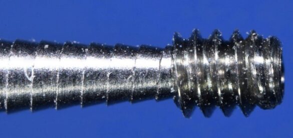
When the appointment was successfully completed I felt the issue with the difficulty in retrieving the fragment was secondary to the distorted top thread, that was until I completed the focus stacked magnified photos for this report. The image below shows the last most apical thread of the fragment literally wiped down to the base of the threads. I’m positive this occurred because this abutment was able to thread into the implant past the threading in the implant and hit the non threaded predrill bore which is 1.25mm in diameter for this M1.6 thread. The only way this happened is if the abutment could seat deeper into the implant as would be the case with an NC abutment.


